1192
Views & Citations192
Likes & Shares
Dermatophytoses
is one of the most frequent human skin diseases of medical importance. Antimicrobial efficacy of Vernonia amygdalina leaves
extracts on some human dermatophytes was studied among fifty (50) selected
Almajiri school children with signs of ringworm, aged between 5 to 10 years and
above in Bauchi metropolis. Aqueous and ethanolic extracts of the leaves were
screened for alkaloids, anthraquinone, cardenolide, flavonoids, phenols,
phlobatannins, saponins, steroids, tannins and terpenoids.
Antifungal activity of the extracts was tested by agar well diffusion method
and Minimum Inhibitory Concentration (MIC) determined. The leave extracts
revealed the presence of all the phytochemicals with the exception of phenol. The disease
is more common (56.5%) in children within 6 to 10 years. All the affected
children had two or more spots on their scalp, indicating presence of Tinea
capitis (scalp ringworm). Microsporum species was
the most frequent (47.8%) dermatophyte isolated, followed by Trichophyton species (23.9%). The zones of inhibition exhibited by
the extracts against the fungal isolates was found within the range of 10.20 to
22.50 mm and varies with the concentration of the extract. Highest MIC value of 65.10mg/ml was found against Epidermophyton and the least 57.50
mg/ml was obtained on Microsporum. These results revealed that the extracts had
significant antimicrobial efficacy against the fungal isolates tested and can
be a cheap source of bioactive materials for the production of anti-dermatophyte
drugs.
Keywords: Dermatophytes, Ringworm, Vernonia amygdalina, Epidermophyton, Microsporum,
Trichophyton
INTRODUCTION
Dermatophytoses is a fungal infection widely
distributed all over the world with various degrees. Many species of these
fungi have been isolated from animals, but a few zoophilic are responsible for
the majority of the cases [1]. The pathological importance of dermatophytes is
associated with contagiousness among the subjects, high cost of treatment,
difficulty of control and the public health outcomes [2].
Dermatophytes are the commonest fungal agents
causing skin diseases. Ringworm is caused by mould fungi of the genera Microsporum, Trichophyton and Epidermophyton.
The location involves are usually the surface of the body (Tinea
corporis), the scalp (Tinea capitis), the foot and the nails (Tinea
unguium or onychomycoses). The fungus settles on the skin, germinates and
forms a mass of branching hyphae which grows out radially to produce circular
lesion [3]. Scalp ringworm (Tinea capitis)
is a superficial fungal infection of the scalp. It is most common in children
4-12 years of age, especially those of black decent [4] and involves red Itchy
patches on the scalp leaving bald spots. It can be persistent and contagious,
almost to the point of epidemic; however, it
Ringworm can pass from one person to another by
direct skin to skin contact or by contact with contaminated items such as
clothes, combs and bathrooms or pool surfaces. Ringworm can also be acquired
from pets that carry the fungus. The first sign of ringworm of the scalp may be
dandruff-like flakes appearing on the hair, round or bald patches. The skin may
feel itchy, showing red and peeling. The rash may gradually spread all over a
large area if prolonged or untreated. Once the hair is infected, it becomes
brittle and breaks off near the root leading to bald spots. Dermatologic lesion
is similar and suspected infection in human should be confirmed by culture to
identify the source of dermatophyte infection [5]. Ringworm is very common
among the less privileged school-age children especially the Almajiris due to
overcrowding, lack of washing clothes, bathing, adequate and beddings. The
Almajiri/Tsangaya system of education is a traditional qur’anic study where
parent give out their sons usually at the age of five to seven and above years
to an individual qur’anic scholar for learning the recitation and memorization
of the holy Qur’an.
Vernonia amygdalina belongs to the plant family
compositae. It is a small shrub that grows
typically grows to a height of 2 to 5 m in tropical Africa. The leaves are petiolate, elliptical and up to 20 cm long and about 6 mm in diameter with rough bark. Vernonia
amygdalina is commonly called bitter leaf in English because of its bitter
taste. In Nigeria, the Hausa calls it Shuwaka, Igbo, Onugbo and Yoruba Ewuro [6]. The leaves are green with a characteristic odour and
bitter taste. It does not produce seeds and has to be distributed or propagated
through stem cutting [4]. It grows under a
range of ecological zones in Africa with about 200 species and produces a lager
mass of forage and it is drought tolerant. It is mainly used for human
consumption and has to be washed to remove the bitter taste. Its bitter taste
is due to anti-nutritional factors such as alkaloids, saponins, tannins and
glycosides. It stimulates the digestive system as well as reduces fever [7]. Vernonia was named after a 17th Century English botanist and plant
collector in North American, Vernon [8].
The plant curative and therapeutic properties
raised over several others vegetables or culinary leaves people make use of in
Nigeria [6]. Its goodness for a healthy body devoid of several diseases like
diabetes and high blood cholesterol problem has also been substantiated by
several research studies both within Nigeria and other African countries where
the plant can be found [9]. The plant roots have been
used for gingivitis and toothache due to its proven antimicrobial activity
[10]. Many herbalists prescribe aqueous extracts for their patients as
treatment for anaemia, nausea, diabetes, loss of appetite, dysentery and other
gastrointestinal track problems. V. amygdalina extracts have also been
reported to help suppress, delay, or kill cancerous cells [11]. However,
extract of bitter leaf had been reported to exert antibiotic action against
drug resistant microorganisms and possess antioxidant, anticancer, antiviral,
anti-helminthic and anti-inflammatory activities [8].
The leaves and bark in local medicine are used as purgative, against menstrual
pain and wound dressing [12].
Ringworm was observed to be common especially
among less privileged children; suffering from poor parenting associated with
personal hygiene and effective health care delivery. In essence, this work will
create awareness on the usefulness and medicinal value of V. amygdalina
as an alternative and affordable to synthetic therapeutic drug by testing it
efficacy on ringworm cases among children. Most of
the previous studies on the antimicrobial efficacy of V. amygdalina extracts
[10,13,14] focused on bacterial infections. Hence the need for this study on
dermatophytes. In Nigeria, the research for new and alternative drugs is on
course, so the present study was designed to evaluate the phytochemical and in vitro anti-dermatophyte activity of Vernonia leaf extracts on
fungal isolates from community cases of ringworm infections.
MATERIALS AND
METHODS
Collection and
preparation of Vernonia leaf samples
Fresh leaf samples of V.
amygdalina were obtained from Fadama garden behind state secretariat,
Bauchi. The plant was identified by a Botanist in the Department of Biological
sciences, Abubakar Tafawa Balewa University (ATBU), Bauchi, Nigeria. The leaves
were aseptically washed, air-dried and grinded into fine powder with pestle and
mortar, and then finally stored in polythene bags until used for ethanolic and
aqueous extractions.
Preparation of cold ethanolic and
aqueous extracts
20 g of the grounded powder of the leaves
material was introduced into a conical flask and 200 ml of absolute ethanol and
distilled water was then added, respectively. The
extraction was carried out at room temperature for 24 h for the aqueous extract
and 72 h for the ethanolic extract. The extract was decanted and filtered with a Whatman No. 1 filter paper
(110 mm). The filtrate obtained was evaporated to dryness at 45°C, and the
obtained residue was discarded as described by Newton et al. [15]. The extract stock solution was filter-sterilized, then stored in
sterile capped tubes in refrigerator at 4°C before use.
Phytochemical screening of the Vernonia leaves
extracts
Phytochemical screening was done in
order to detect the presence of following bioactive compounds: alkaloids, anthraquinone, cardenolide, flavonoids, phenols, phlobatannins,
saponins, steroids, tannins and terpenoids using the methods
described by Wazis et al. [16] and
Sofowora [17].
Alkaloids: A 3 mm of the ethanolic and aqueous extracts was
stirred with 5 ml of 1% HCL on a steam bath for twenty minutes. The solution
obtained was cooled and filtered and few drops of Mayer’s reagent/picric acid
were added to the filtrate. A cream precipitate indicated the presence of
alkaloid.
Anthraquinone:
A 0.5 g of the plant extract was mixed with 10
ml of aqueous H2SO4 and then filtered while hot, the filtrate
was shaked with 5 ml of benzene, the benzene layer separated and
half its own volume of 10% ammonia solution was then added. The presence
of violet or red coloration in the ammonical (lower) phase was
taken as positive combined anthraquinone.
Cardenolide: A 0.5 g of the
plant extract was dissolved in 2 ml of glacial acetic acid containing a
drop of ferric chloride solution. This was then underlayed with 1ml
of concentrated tetraoxosulphate (VI) acid. Appearance of a brown
at the interphase showed the presence of digitoxose sugar characteristic
of cardenolide.
Flavonoids: A volume of 3 mm of the
ethanolic and aqueous extract was added to a volume of 1 ml of 10% sodium
hydroxide. A yellow coloration indicated the presence of flavonoids.
Glycosides: A 2
ml of chloroform was added to a volume of 3 ml of the ethanolic and aqueous
extract. Dilute sulphuric acid was carefully added to form a lower layer. A
reddish brown colour at interface indicated the presence of a steroidal ring.
Phenolics: Two drops of 5% ferric
chloride were added to 5 ml of the ethanolic and aqueous extracts in a test
tube. A greenish precipitate was observed as positive for phenolics.
Phlobatannins: A 1%
hydrochloric acid was added to a volume of 1 ml of the ethanolic and aqueous
extracts. A red precipitate was regarded as the presence of phlobatannins.
Saponins: 2 ml of the aqueous and
ethanolic extracts in a test tube was shaken for 2 min. Frothing which
persisted on shaking was taken as evidence for the presence of saponins.
Steroids: To a volume of 1 ml of
the extracts, five drops of concentrated tetra-oxoosulphate VI acid (H2SO4) was added. Red coloration indicated the
presence of steroids.
Tannins: A volume of 1 ml of
freshly prepared 10% potassium hydroxide was added to a volume of 1 ml of the
ethanolic extracts and aqueous extracts. The presence of a dirty white
precipitate was considered as indication of tannins.
Terpenoids: A 10 ml of
extracts was mixed with 2 ml Chloroform and 3 ml of concentrated H2SO4
was carefully added to form a layer. A reddish brown coloration of the
interface formed indicating the presence of terpenoids.
Sample collection
The specimens were
collected from different parts of the body or scalp of 50 randomly selected
school-age children in Almajiri houses (Tsangaya) within Yelwa and Gwallameji
area of Bauchi metropolis. A new surgical blade was used for each individual
child. The specimens were collected by gently scraping affected spots into
clean sheets of paper which were then transferred into sterile containers that
had been properly labelled with respect to each individual’s data; these were
brought to the laboratory for inoculation. Informed consent
of the child and their teachers was obtained before the sample collection. The
participation was open and voluntary.
Microscopic
identification
A drop of potassium hydroxide solution was
placed on a clean sterile glass slide and small clean pieces of the specimen
was transferred to the drop of potassium hydroxide and covered with a cover
slip. The preparation was then examined using 10x and 40x objectives with the
condenser iris diaphragm closed sufficiently to give good contrast for the
presence of branching hyphae and rounded anthrospores.
Culture
methods
The specimen
was inoculated onto Sabouraud
dextrose agar (SDA) media (Oxoid, UK) and incubated at
room temperature for four days, after which it was sub-cultured. The isolates were finally stored on SDA
slants in the refrigerator at 4°C prior to use, as described by Chander [18].
Screening
for antifungal activity of the extracts
The ethanolic extract of the Vernonia leaves was
applied on the fungal isolates Epidermophyton,
Trichophyton and Microsporum species using agar diffusion as described
by Newton et al. [15]. The fungal isolates were allowed to grow on a Sabouraud
dextrose agar (SDA) (Oxoid, UK) at 25℃ until they sporulated. The fungal spores
were harvested after sporulation by pouring a mixture of sterile glycerol and
distilled water to the surface of the plate and later scraped the spores with a
sterile glass rod. 100 µl of the standardized fungal spore suspension was
evenly spread on SDA media. Wells were then bored into the agar media using a
sterile 6 mm cork borer and then carefully filled with the extracts. The plates
were allowed to stand on the laboratory bench for 1 hour to allow for proper
diffusion of the extract into the media. Dimethyl Sulfoxide (DMSO) was used as
a negative control and Griseofulvin was used as a positive control. The plates
were incubated at 25℃ for 96 h and later examined for zones of inhibition.
Determination of minimum inhibitory
concentration (MIC)
The MIC of the aqueous extract of Vernonia leaves was
estimated using the methods of Rebell and Taplin [19]. Two-fold dilutions of the extract
was prepared and 2 ml aliquots of different concentrations of the solution were
added to 18 ml of pre-sterilized molten SDA for fungi at 40℃ to give final
concentration solutions of 10 mg/ml. The medium was then poured into sterile
Petri dishes and allowed to set. The surface of the medium was allowed to
air-dry under laminar flow, then inoculated with the old fungal cultures. The
plates were later incubated at 25℃ for 3 days and later observed for the
presence or absence of growth. The MIC was taken as the lowest concentration
that prevented the growth of the test isolates.
RESULTS
Bioactive
constituents of Vernonia amygdalina leave extracts
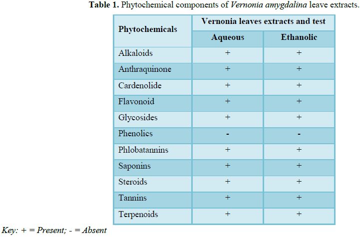
Phytochemical
screening of the Vernonia leaves extracts in this study (Table 1) revealed the presence of alkaloids, anthraquinone,
cardenolide, flavonoids,
phlobatannins, saponins, steroids, tannins and terpenoids,
with the exception of phenols, in both the aqueous and ethanolic
extracts.
Children
affected with dermatophytoses
These studies
observe the prevalence of dermatophytes among the Almajiri children in relation
to their age (Table 2). It was found
that the disease is most prevalent (56.5%) in children within 6 to 10 years,
followed by those above. However, some of the children were found as young as 4
to 5 years and uncircumcised. All the affected children have two or more spots
on their scalp, indicating presence of Tinea capitis (scalp ringworm).
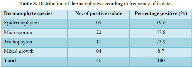
This study also found that Microsporum species was the most frequent (47.8%) dermatophyte
isolated, followed by Trichophyton
species (23.9%) (Table 3).
Antidermatophyte
activity of V. amygdalina leaves extracts
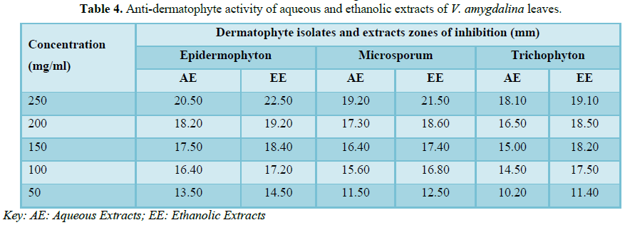
Antifungal efficacy of
the Vernonia leaves extracts was tested against the isolates of Epidermophyton Microsporum and Trichophyton
species. The
average of the zones of inhibition for each extract was then calculated (Table 4). The zones of inhibition
exhibited by the extracts against the fungal isolates was found within the
range of 10.20 to 22.50 mm and varies with the concentration of the extract.
The diameter zone of inhibition decrease with decrease in the concentration in
both the extracts, where the highest zones correspond to highest concentration.
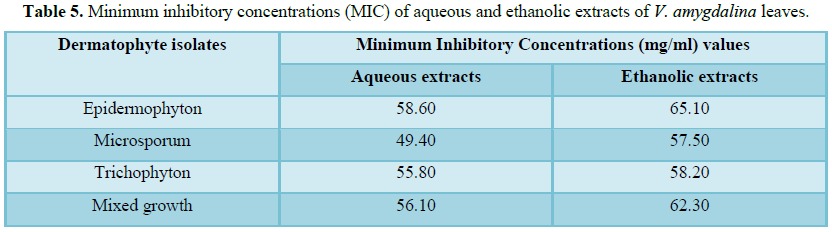
The minimum
inhibitory concentrations (MIC) of the ethanolic extracts of Vernonia amygdalina
(mg/ml) was also analyzed in this study (Table
5). The highest MIC value of 65.10 mg/ml was found (P<0.05) against Epidermophyton and least MIC
value of 57.50 mg/ml was obtained against Microsporum.
DISCUSSION
Phytochemical
substances have profound antimicrobial activity on various infectious agents
through different mode of actions. The metabolites are found to be biologically
active and play vital roles in the therapeutic activity of medicinal plants
with specific action on human body. Previous studies by Imaga
and Bamigbetan [7] on Vernonia amygdalina
leaves extracts revealed also confirmed the presence of these compounds.
However, phenols was found in Vernonia leaves in this study, the analysis of ethanolic extract of the Vernonia leaves by Alara et al. [2] revealed the presence of these compound but
with absence of anthraquinone. Among these phytochemicals, alkaloids are having
useful effect on humans, as it serves as a component of powerful pain relievers
[10]. The present study observed the efficacy Vernonia leaves extracts against fungi.
Dermatophytoses
is a fungal infection commonly affecting school age children especially the
less privileged. Scalp
ringworm is highly contagious especially among children [3]. The Almajiri children in northern Nigeria suffered from total
negligence due to poor parental background, non-chalant attitudes and religious
misconception of some parents. The children are left without proper daycare
needs such as beddings, bathing and washing, due to their large number under a
single Qur’anic scholar and the children had to cater for all the daily needs
by roaming about the streets, begging for food and money. They sometimes scout
for junks along waterways and refuse dumps to sell. These ill-health conditions
exposed them to some fungal pathogens, including the dermatophytes. Microsporum canis was found to be the most common
fungal agent associated with dermatophytoses and accounting for up to 70% of
the infection in a similar study by Bokhari [1].
The rise in the
prevalence of side effects of many synthetic antimicrobial agents and emergence
of multidrug resistant fungi encouraged research for plant-based drugs of
therapeutic potentials. Vernonia amygdalina was reported to have
such potential of high medicinal value [2]. In this study, aqueous extracts of
this plant showed high antifungal activity against the isolates of Epidermophyton, Microsporum and Trichophyton
species at 58.60, 49.40 and 55.80 mg/ml concentrations
respectively. While for ethanolic extracts had the highest respective
concentrations and activity (P<0.05) as 65.10,
57.50 and 58.20 against the isolates. The ethanolic extracts of the
leaves show more antifungal properties than the aqueous extracts (P<0.05),
which may due to the solvents used. Organic extract was found to be more active
than water extract due to the better solubility of the active components in
organic solvents [5].
The extracts
however, were active against fungi of medical importance. The presence of alkaloids, anthraquinone, cardenolide, flavonoids, phlobatannins,
saponins, steroids, tannins and terpenoids in the
extracts of V. amygdalina in this study may explain the reason for its
antifungal activities as the antimicrobial properties of most of these
phytochemicals have been previously reported [5,6].
CONCLUSION
This study
showed that the extracts from these leaves revealed significant antifungal
activities (P<0.05) on all the fungal isolates tested and might be source of
active ingredients for the synthesis of antibiotics. The phytochemical
components are quite promising and have strongly indicated the anti-dermatophyte
efficacy of the plant leaves. As the findings of this study compared favorably
with previous studies on fungal infections, the plant holds great promise for
use as antimicrobial agent. The efficacy gives impetus to the use of these
plants in meeting health care needs of infected children. Further studies are
required to characterize the Vernonia leaves active components by molecular
techniques of these plants. Cytotoxicity levels should be evaluated using
laboratory animals so that these extracts can be formulated into tablets and
creams that can be used to treat dermatophytoses and other related fungal
infections. Other methods of extraction should be tried to determine the best
method for optimal yield of the bioactive constituents.
1. Bokhari FM (2009) Antifungal activity of some
medicinal plants used in Jeddah, Saudi Arabia. Mycopathology 7: 51-57.
2. Alara OR, Abdurrahman MN, Ukaegbu CI, Kabbashi NA
(2018) Extraction and characterization of bioactive compounds in Vernonia
amygdalina leaf ethanolic extract comparing Soxhlet and microwave-assisted
extraction techniques. J Taibah Univ Sci 13: 122-126.
3. Cheesbrough M (2012) District laboratory practice
in tropical countries. Part two (2), Cambridge University Press: Cambridge, UK,
pp: 67-81.
4. Oboh FOJ, Masodje HI (2009) Nutritional and
antimicrobial properties of Vernonia amygdalina leaves. Int J Biomed
Health Sci 5: 51-56.
5. Inusa A, Sunusi SB, Linatoc AC, Mainasara MM, Awawu
JJ (2018) Phytochemical analysis and antimicrobial activity of bitter leaf (Vernonia
amygdalina) collected from Lapai, Niger state, Nigeria on some selected
pathogenic microorganisms Sci World J 13: 15-18.
6. Adetunji CO, Olaniyi OO, Ogunkunle ATJ (2013)
Bacterial activity of crude extracts of Vernonia amygdalina on clinical
isolates. J Microbiol Antimicrob 5: 60-64.
7. Imaga NOA, Bamigbetan DO (2013) In vivo biochemical
assessment of aqueous extracts of Vernonia amygdalina (bitter leaf). Int
J Nutr Metab 5: 22-27.
8. Asuzu CU (2018) Bitter herbs of eastern Nigeria (Gongronema
latifolium, Vernonia amygdalina and Vitex doniana): A review.
Afr J Tradit Complement Alternat Med 15: 47-56.
9. Yeap SK, Ho WY, Beh BK, Liang LS, Huynh K, et al.
(2010) Vernonia amygdalina, an ethnoveterinary and ethnomedical used
green vegetable with multiple bioactivities. J Med Plants Res 4: 2787-2812.
10. Ghamba PE, Balla H, Goje LJ, Halidu A, Dauda MD
(2014) In vitro antimicrobial activities of Vernonia amygdalina
on selected clinical isolates. Int J Curr Microbiol Appl Sci 3: 1103-1113.
11. Luke UO, Ebong PE, Eyong EU, Robert AE, Ufot SU, et
al. (2013) Effect of ethanolic root and twig extracts of Vernonia amygdalina
(Etidot) on liver function parameters of streptozotocin induced hyperglycemic
and normal Wistar rats. Eur Sci J 9: 1857-7881.
12. Uhegbu FO, Ogbuehi KJ (2004) Effect of aqueous
extract (crude) of leaves of Vernonia amygdalina (Del) on blood glucose,
serum albumin and cholesterol levels in diabetic albino rats. Glob J Pure Appl
Sci 10: 189-194.
13. Ibrahim TA, Ajala L, Adetuyi FO, Jude-Ojei B (2009)
Assessment of the antibacterial activity of Vernonia amygdalina and Ocimum
gratissimum leaves on selected foodborne pathogens. EJEAFC 8: 1212-1218.
14. Oshim IO, Desmond CO, Nwobu RA, Ezugwu UM, Urama EU
(2016) Kinetics of minimum inhibitory concentration, minimum bactericidal
concentration and minimum fungicidal concentration of Vernonia amygdalina
(bitter leaf) on microorganisms isolated from wound infections. Int J Surg Res
5: 8-14.
15. Newton SM, Lau C, Gurcha SS, Besra GS, Wright CW
(2002) The evaluation of forty-three plant species for in vitro anti-mycobacterial
activities isolation. Int J Curr Microbiol Appl Sci 3: 1103-1113.
16. Wazis CH, Timothy SY, Zakama SG, Balla HJ, Maspalma
JD (2013) Phytochemical screening and purgative activity of ethanolic extracts
of Vernonia amygdalina Del. leaf. Int J Res Ayurveda Pharm 4: 46-49.
17. Sofowora EA. (1993) Medicinal plant and traditional
medicine in Africa. 2nd Edn. Spectrum Book: Ibadan, Nigeria, pp:
261-265.
18. Chander J (2002) A Textbook of Medical Mycology. 2nd
Edn, pp: 212-227
19. Rebell G, Taplin D (2008) Dermatophytes: Their
recognition and identification. University of Miami Press: USA, pp: 67-68.
-
 Table 1
 -
Table 2
-
Table 3
-
Table 4
-
Table 5

QUICK LINKS
- SUBMIT MANUSCRIPT
- RECOMMEND THE JOURNAL
-
SUBSCRIBE FOR ALERTS
RELATED JOURNALS
- Journal of Nursing and Occupational Health (ISSN: 2640-0845)
- Chemotherapy Research Journal (ISSN:2642-0236)
- International Journal of Diabetes (ISSN: 2644-3031)
- Advance Research on Endocrinology and Metabolism (ISSN: 2689-8209)
- Journal of Otolaryngology and Neurotology Research(ISSN:2641-6956)
- Journal of Allergy Research (ISSN:2642-326X)
- Journal of Pathology and Toxicology Research
")
")


